
Speaker: DenTrends
Session Type: Radiology in Implantology
Oral & Maxillofacial Surgeon, Hanyang University Hospital
In this DenTalk session, Professor Park Chang Jo shares practical insights into the critical role of radiology in modern implantology. With clarity and simplicity, he bridges foundational concepts with advanced digital workflows, helping clinicians understand why radiology is not optional—but essential.
Why Radiology Matters in Implant Dentistry
Dentistry evolves rapidly, but strong fundamentals remain the backbone of safe and predictable implant treatment. Radiology plays a decisive role in:
- Accurate diagnosis
- Individualized treatment planning
- Avoiding anatomical complications
- Evaluating surgical outcomes
Every implant case begins and ends with imaging.
Imaging Modalities Explained in the Session
CBCT (Cone Beam Computed Tomography)
CBCT stands at the center of digital implantology, offering:
- High-resolution 3D images
- Precise visualization of bone, sinuses, and nerves
- Reduced radiation compared to conventional CT
- Essential data for digitally guided implant surgery
Without CBCT, true digital implant workflows are impossible.
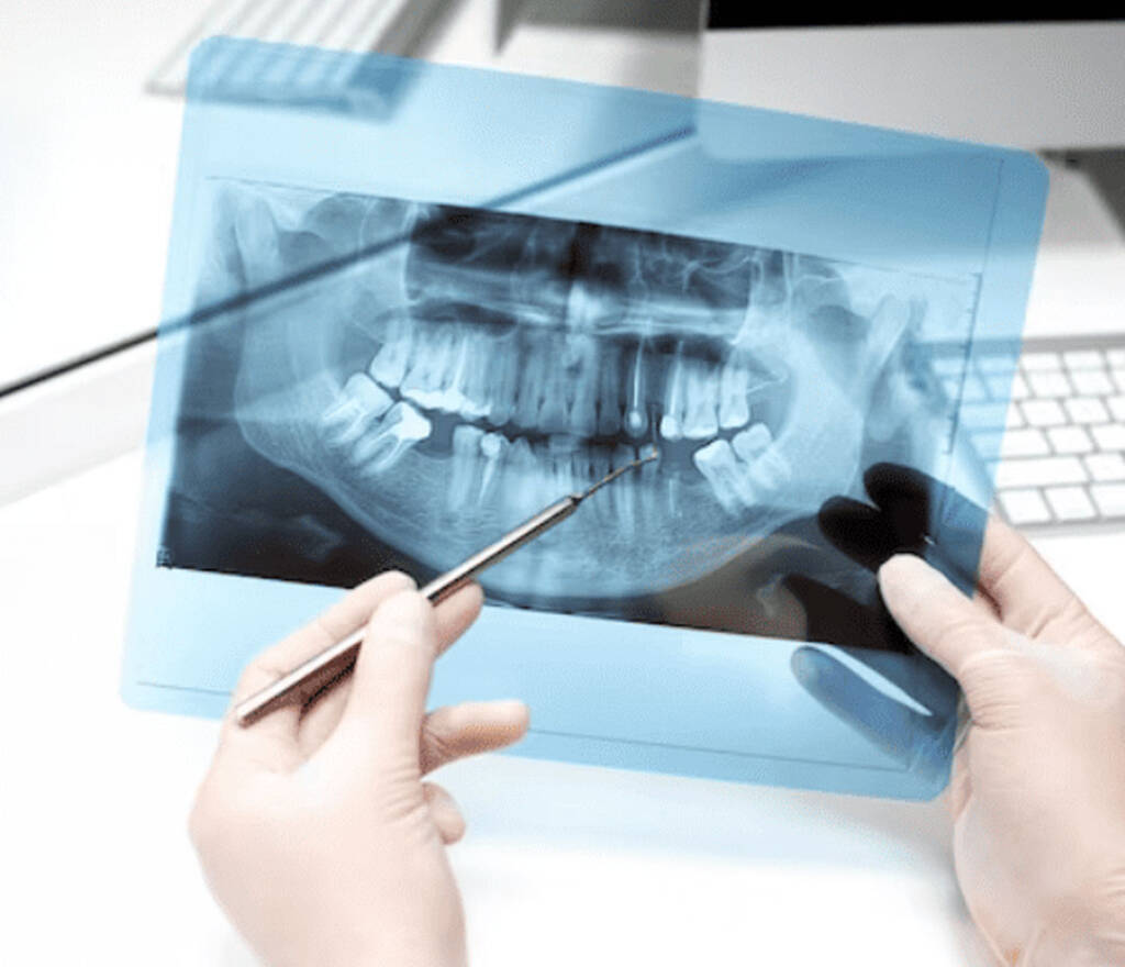
Panoramic Radiology
Still the first-line imaging tool worldwide, panoramic radiographs provide:
- Broad overview of jaws and teeth
- Identification of anatomical landmarks
- Quick screening for pathology
- Guidance on whether CBCT is required
Despite digital advances, panoramas remain indispensable in daily practice.
Periapical Radiology
Used for high-detail evaluation of localized areas, periapical X-rays help assess:
- Implant-to-bone interface
- Implant-to-tooth relationships
- Fine structural details
Best suited as a complementary tool rather than a standalone solution.
Clinical Insight from the Session
Through complex implant cases, Professor Park demonstrates how CBCT enables:
- Multi-stage treatment planning
- Sinus lift evaluation
- Guided bone regeneration assessment
- Post-operative outcome monitoring
These procedures cannot be planned accurately using panoramic images alone.
From Analog to Digital Implant Surgery
The session highlights the evolution from:
- Manual tracing on panoramic films
- Radiologic stents and surgical templates
- Fully digital planning
- CBCT-based guided implant systems
Modern guided surgery is essentially a refined digital version of early analog principles.
Radiation Safety Reassurance
A key takeaway for clinicians and patients alike:
- Dental radiography doses are extremely low
- CBCT and panoramic imaging are well within safe limits
- Everyday activities (like long-haul flights) expose people to more radiation
Radiologic imaging in dentistry is safe, controlled, and justified.
Key Takeaways from This DenTalk
- CBCT is the backbone of digital implantology
- Panoramic radiology remains a vital screening tool
- Periapical radiographs add precision in localized assessment
- Radiology ensures safety, accuracy, and long-term implant success
- Careful interpretation of images prevents complications and missed pathology
DenTalk Conclusion
This DenTalk session sets a strong foundation for implant learners, reinforcing that mastery begins with understanding radiology. Professor Park encourages continuous learning and reminds clinicians that technology enhances skill—but fundamentals ensure success.